Indiana University School of Medicine – Bloomington

The Segal Lab

Mechanisms of Cell–Microenvironmental Interactions in Cancer

We use quantitative, high-resolution imaging in 3D culture and zebrafish avatar models to ask how the tissue microenvironment drives cancer cell plasticity — and how that plasticity enables survival, adaptation, and therapy resistance.

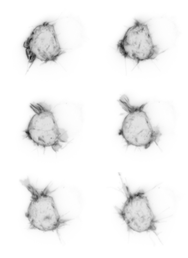
  • Live imaging of a cell expressing Caveolin-1-GFP
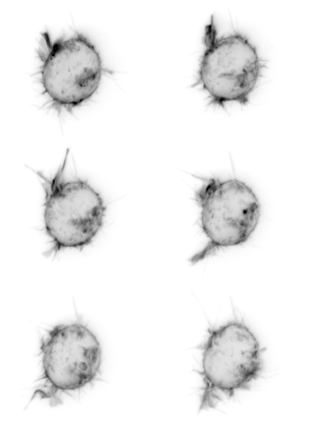
  • Live imaging of a cell expressing an active PI3K sensor
  • Live-cell imaging of a cancer cell with membrane protrusions
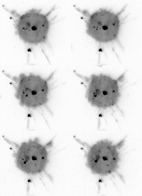
  • Live-cell imaging of a cancer cell
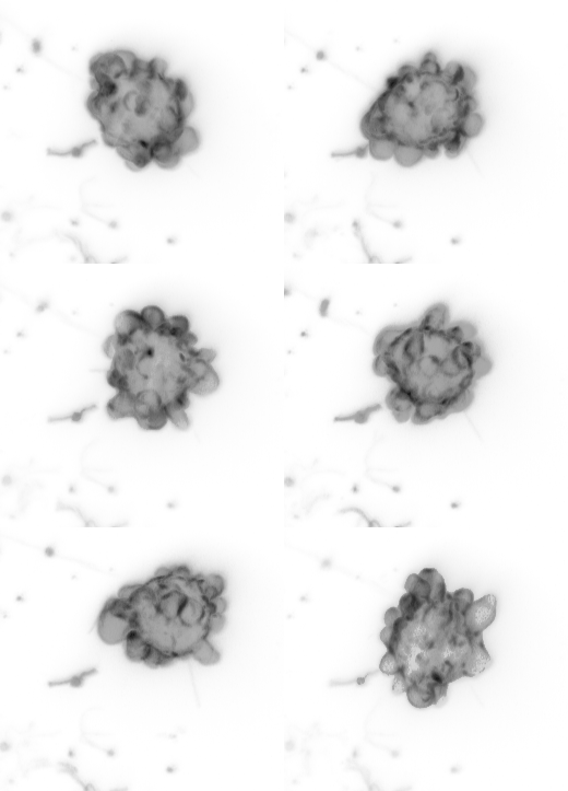
  • Live-cell imaging of a cancer cell

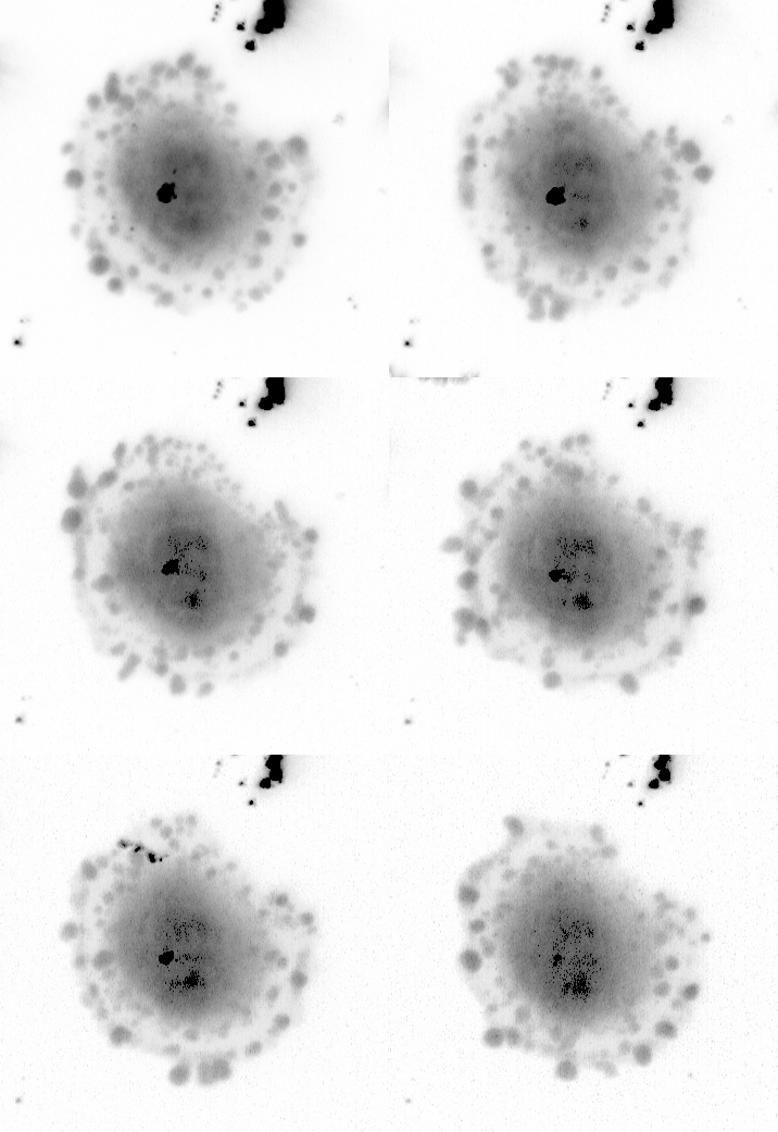
Ewing Sarcoma cells display dynamics of active PI3K signaling organization

Research

Mechanisms of Cell–Microenvironmental Interactions in Cancer

Cancer cell plasticity is a key driver of disease progression, enabling metastasis and resistance to therapy — yet the mechanisms by which cancer cells sense and adapt to external cues remain largely unknown. This gap is especially critical in pediatric cancers, where a low mutational burden beyond a single driver oncogene often contrasts sharply with the variability seen in patient outcomes, pointing to non-genetic, microenvironment-driven mechanisms of adaptation.

Our work centers on Ewing Sarcoma (EwS), a pediatric cancer of bone and soft tissue, as a prototype for studying these questions. Traditional multi-omics approaches capture transcriptional states but often miss the cell-biological processes — membrane dynamics, cytoskeletal reorganization, spatial compartmentalization of signaling — that actually drive cell function. We address this by developing and applying quantitative live-imaging approaches that resolve cancer cell behavior directly, in physiologically relevant contexts.

Zebrafish avatars

High-resolution, high-throughput live imaging of human cancer cells xenografted into distinct tissue microenvironments in a living vertebrate.

3D culture systems

Engineered extracellular matrix environments to dissect how specific cell–ECM interactions shape cell state and signaling.

Quantitative imaging

Custom analytical pipelines that turn subcellular imaging data into quantitative, morphology- and signaling-based readouts of cell state.

Current research directions

01

Chromatin remodeling and EWSR1⁠:⁠FLI1 activity

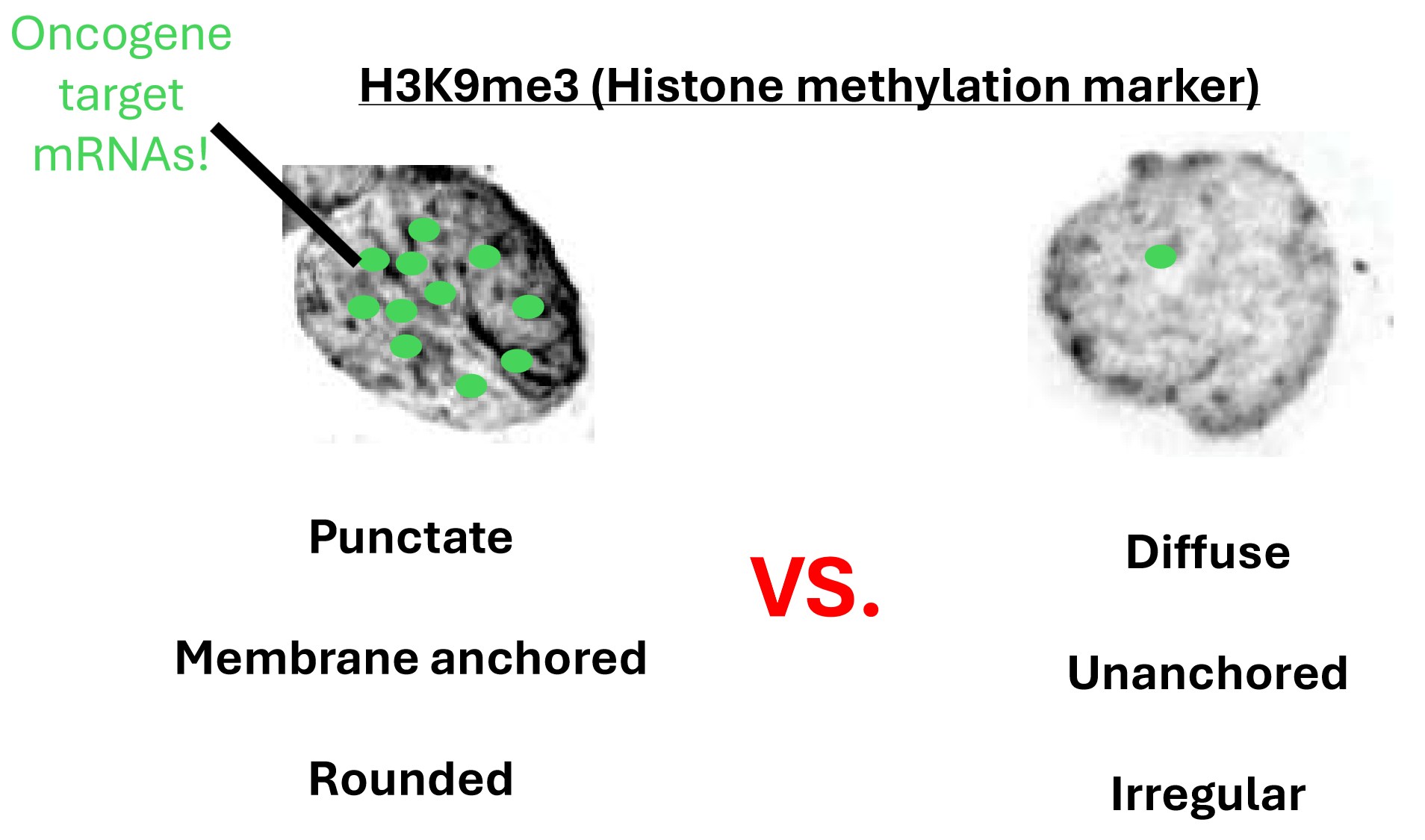
H3K9me3 histone methylation in two nuclear states: punctate, membrane-anchored and rounded versus diffuse, unanchored and irregular, with oncogene target mRNAs marked

We are asking how chromatin state contributes to Ewing Sarcoma plasticity, beginning with how EWSR1:FLI1 (EF1) activity differs across microenvironments. Live imaging of histones and histone acetylation markers lets us follow nuclear changes as cells shift between environments. Building on this, we are designing transcriptional reporters of EF1 targets to visualize EF1 activity states in vivo, in zebrafish, across different tissue contexts.
02

ECM cues and cytoskeletal regulation

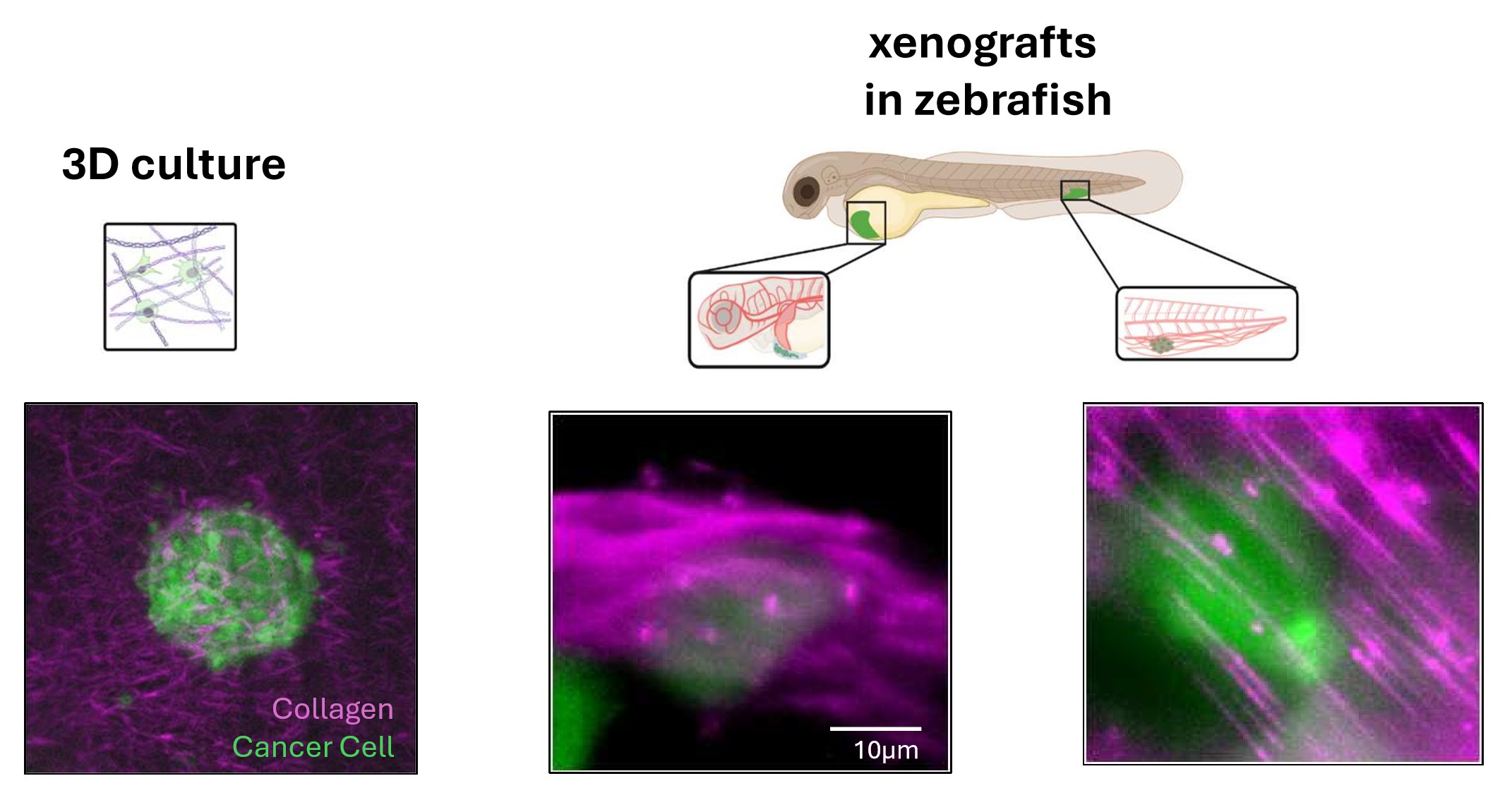
Cancer cells and collagen in 3D culture and in zebrafish xenografts at two tissue sites

Changes in matrix composition and density alter cell shape, which shifts the polarization of Rac and with it the balance between cell migration and proliferation — a balance relevant to Ewing Sarcoma progression. We are perturbing Rac and Dia to dissect this pathway, defining the cytoskeletal regulators that translate ECM cues into cell state.

03

Membrane fluidity and cell state

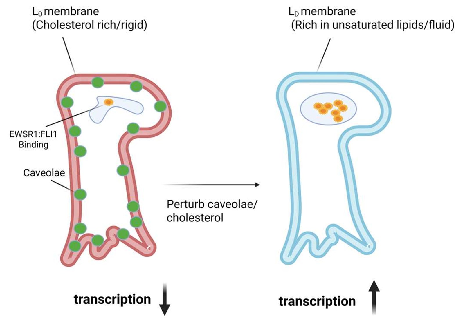
Model: a cholesterol-rich, rigid membrane with caveolae reduces EWSR1:FLI1 transcription; perturbing caveolae or cholesterol yields a fluid membrane and increased transcription

We are profiling differences in membrane composition between CD99-high and CD99-low Ewing Sarcoma states, using membrane dyes followed by lipidomics. Differences we identify are genetically perturbed to test their effect on cell phenotype, nominating membrane-regulating pathways that could be targeted therapeutically.

04

Adhesion and tissue-specific adaptation

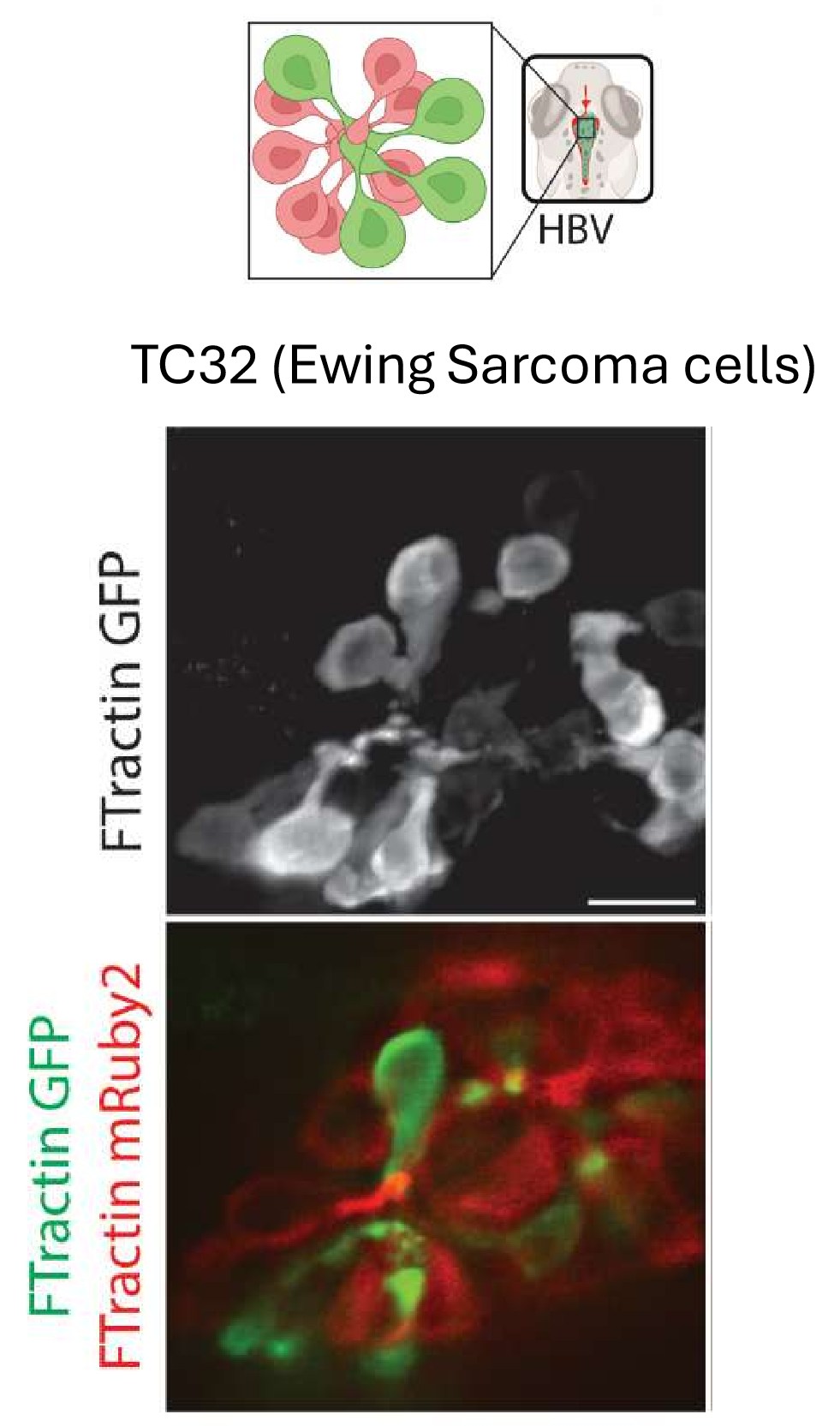
Pseudorosette formation by TC32 Ewing Sarcoma cells in the zebrafish hindbrain ventricle, imaged with FTractin-GFP and FTractin-mRuby2

EwS cells form pseudorosettes — radially organized architectures seen clinically and in development — but only in specific tissue microenvironments, such as the zebrafish hindbrain ventricle. We are identifying the adhesion molecules that drive this tissue-specific self-organization, combining mosaic cell labeling with long-term light-sheet imaging to resolve their functional roles.

Together, these projects will define how microenvironmental cues orchestrate cell state transitions in cancer — building a foundation that extends beyond Ewing Sarcoma, and toward zebrafish patient-derived avatars for precision, context-aware cancer therapy.

People

Lab Members

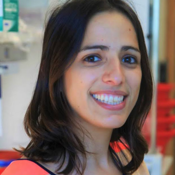
Dagan Segal, Ph.D.

Dagan Segal, Ph.D.

Principal Investigator · Assistant Professor

The Segal Lab is opening at Indiana University School of Medicine – Bloomington in 2026. We are actively recruiting our founding lab members — see Join Us for opportunities.

Join Us

Join the Lab

We are building our founding team and welcome inquiries from prospective graduate students, postdoctoral fellows, research associates, and undergraduate researchers who are excited about quantitative imaging, cancer cell biology, and in vivo model systems.

Publications

Selected Publications

  1. Haynes KA, Adebowale K, Akpa BS, Bader JS, Bansal T, Breunig J, Brosh R, Bugaj LJ, Burslem GM, Chadha A, Das M, Deans T, DeForest CA, Dooling LJ, Gartner Z, Heiser LM, Hughes A, Kemp ML, Korolev KS, Lin C, Lionnet T, Lu X, McGuigan AP, Mirzaei HR, Nirmal AJ, Pasca di Magliano M, Poulopoulos A, Rai K, Reyes J, Segal D, Sewell-Loftin MK, Shmeleva EV, Stevens A, Subramanian S, Tlsty T, Totonchy J, Vlahos A, Wang P, Zervantonakis IK, Zhao Y, Chuang JH. (2026) Integrating Technologies for Synthetic, Systems, and Spatial Approaches in Cancer Research. ACS Synthetic Biology (Perspective). doi:10.1021/acssynbio.6c00339
  2. Daetwyler S, Mazloom-Farsibaf H, Zhou FY, Segal D, Sapoznik E, Westcott JM, Brekken RA, Danuser G, Fiolka RP. (2025) Imaging of cellular dynamics in vitro and in situ: from whole organisms to sub-cellular imaging with self-driving, multiscale microscopy. Nature Methods 22(3), 569–578.
  3. Segal D, Wang X, Mazloom-Farsibaf H, Rajendran D, Butler E, Chen B, Chang B-J, Ahuja K, Perny A, Bhatt K, Reed DK, Castrillon D, Lee J, Jeffery E, Wang L, Williams NS, Rajaram S, Fiolka RP, Skapek SX, Hon GC, Amatruda J, Danuser G. (2024) Caveolin-1 regulates context-dependent signaling and survival in Ewing Sarcoma. bioRxiv (preprint). doi:10.1101/2024.09.23.614468
  4. Kamalesh K, Segal D, Avinoam O, Schejter ED, Shilo B-Z. (2024) Structured RhoGEF recruitment drives myosin II organization on large exocytotic vesicles. Journal of Cell Science 137(13):jcs.261944.
  5. Segal D, Mazloom-Farsibaf H, Chang B-J, Roudot P, Rajendran D, Fiolka R, Warren M, Amatruda JF, Danuser G. (2022) In vivo 3D profiling of human cancer cell morphotypes in zebrafish. Journal of Cell Biology 221(11):e202109100.
  6. Perez-Castro L, Venkateswaran N, Garcia R, Hao Y-H, Lafita-Navarro MC, Kim J, Segal D, Saponzik E, Chang B-J, Fiolka R, Danuser G, Xu L, Brabletz T, Conacci-Sorrell M. (2022) The AHR target gene Scinderin activates the Wnt pathway by facilitating nuclear translocation of β-catenin. Journal of Cell Science 135(20):jcs260028.
  7. Segal D, Zaritsky A, Schejter ED, Shilo B-Z. (2018) Formation and disassembly of a contractile actomyosin network mediates content release from large secretory vesicles. Journal of Cell Biology 217(5):e201711006.
  8. Segal D. (2017) Live Imaging of Myogenesis in Indirect Flight Muscles in Drosophila. Bio-protocol 7(13):e2377. doi:10.21769/BioProtoc.2377
  9. Segal D, Dhanyasi N, Schejter ED, Shilo B-Z. (2016) Adhesion and Fusion of Muscle Cells are Promoted by Filopodia. Developmental Cell 38, 291–304.
  10. Dhanyasi N, Segal D, Shimoni E, Shinder V, Shilo B-Z, VijayRaghavan K, Schejter ED. (2015) Surface apposition and multiple cell contacts promote myoblast fusion in Drosophila flight muscles. Journal of Cell Biology 211, 191–203.
  11. Kidd MR, Dijkstra PD, Alcott C, Lavee D, Ma J, O'Connel LA, Hofmann HA, Segal D. (2013) Behavioral Ecology and Sociobiology 67:1307–1315.

For a complete publication list, see Google Scholar or PubMed.

News

Lab News

  • 2026

    Welcome to the Segal Lab! We are opening our doors at Indiana University School of Medicine – Bloomington and recruiting our founding team.

Contact

Get in Touch

Prospective students, postdocs, and collaborators are welcome to reach out directly.

Phone
(812) 856-1207
Location
200 Myers Building
Indiana University School of Medicine – Bloomington
Bloomington, Indiana